Anatomy is a branch of biology that is the consideration of the structure of living things. It is a general term that includes human anatomy, animal anatomy (zootomy) and plant anatomy (phytotomy)

Friday, May 30, 2008

Karl Georg Friedrich Rudolf Leuckart (1822-1898)

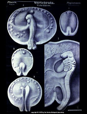
Rudolf Leuckart was considered the "Father of Parasitology" as well as one of the most famous zoologists of the 19th century. He began his career in zoology shortly after earning a M.D. degree from the University of Gottingen when he embarked on a scientific expedition to the North Sea to study marine invertebrates. His careful descriptions of morphologic details gave major support to the newly established field of animal systematics: the idea that evolution can be traced through structural changes. Leuckart was awarded an assistant professorship at age 28 followed in 1869 by a full professorship at the University of Leipzig.

More pictures Here

i will upload allot more pictures from Rudolf Leukart


Thursday, May 29, 2008

Radiology

Radiologic technologists take x rays and administer nonradioactive materials into patients’ bloodstreams for diagnostic purposes.

Radiologic technologists also referred to as radiographers, produce x-ray films (radiographs) of parts of the human body for use in diagnosing medical problems. They prepare patients for radiologic examinations by explaining the procedure, removing jewelry and other articles through which x rays cannot pass, and positioning patients so that the parts of the body can be appropriately radiographed. To prevent unnecessary exposure to radiation, these workers surround the exposed area with radiation protection devices, such as lead shields, or limit the size of the x-ray beam. Radiographers position radiographic equipment at the correct angle and height over the appropriate area of a patient’s body. Using instruments similar to a measuring tape, they may measure the thickness of the section to be radiographed and set controls on the x-ray machine to produce radiographs of the appropriate density, detail, and contrast. They place the x-ray film under the part of the patient’s body to be examined and make the exposure. They then remove the film and develop it

More on Radiology over at Sumerdoc


Wednesday, May 28, 2008

Re-Animator (1985)

I have to post these screen shots from the intro to Re-Animator, that intro is so cool.

Re-Animator (1985) is the first in a series of films based on the H. P. Lovecraft story "Herbert West: Reanimator". It stars Jeffrey Combs as Dr. Herbert West, a medical student who transfers from a school in Europe to the medical school of Miskatonic University to continue developing a formula to revive the dead. His testing of the agent leads to unintended consequences for West and his new roommate, Dan Cain. The movie has since become a cult film, driven by fans of Combs, extreme gore, and successful combination of horror and comedy. It currently has a score of 91% on critic site RottenTomatoes.com.

More pictures at my Flickr Here 

The film is available here Re-Animator


Saturday, May 24, 2008

Govard Bidloo (1649 - 1713)

Govard Bidloo was born in Amsterdam in 1649 and became professor of anatomy at The Hague from 1688 to 1694, when he took the same position at Leyden. He was later appointed the physician of William III of England, who was originally Dutch, until the King's death in 1702. In that year, Bidloo returned to Leyden to take his old position until his death there in 1713. 

Best known as an anatomist, Govard Bidloo's most famous work was his monumental Anatomia humani corporis published in Amsterdam in 1685, containing 107 copperplate engravings. Like so many large and expensive anatomical atlases of the time, the work was not a financial success, and in 1690 he published a Dutch translation entitled, Ontleding des menschelyken lichaams, using the same plates. 

When this edition did not sell well either, Bidloo's publisher sold 300 of the extra printed plates to William Cowper, a noted English anatomist. Cowper published the plates with his own, English language text in Oxford in 1698 under the title, Anatomy of the humane bodies, without mentioning Bidloo or the artists of the original plates. Cowper went so far as to use Bidloo's engraved allegorical title page, amended with an irregular piece of paper lettered: "The anatomy of the humane bodies ...," which fits over the Dutch title (see a comparison here). 

A number of vitriolic exchanges took place between Bidloo and Cowper, including several pamphlets published in each anatomist's defense. Cowper claimed, without much evidence presented, that the plates were not Bidloo's at all, but that they were commissioned by Jan Swammerdam (1637-1680) and that after his death Swammerdam's widow had sold them to Bidloo. 

More Pictures Here


Thursday, May 22, 2008

Bernhard Seigfried Albinus (1697-1770)

Bernhard Siegfried Albinus (i.e. Weiss) was born in Frankfurt an der Oder on February 24, 1697, the son of the physician Bernhard Albinus (1653-1721). He studied in Leyden with such notable medical men as Herman Boerhaave, Johann Jacob Rau, and Govard Bidloo and received further training in Paris. He returned to Leyden in 1721 to teach surgery and anatomy and soon became one of the most well-known anatomists of the eighteenth century. He was especially famous for his studies of bones and muscles and his attempts at improving the accuracy of anatomical illustration. Among his publications were Historia muscolorum hominis (Leyden, 1734), Icones ossium foetus humani (Leyden, 1737), and new editions of the works of Bartholomeo Eustachio and Andreas Vesalius. Bernhard Siegfried Albinus died in Leyden on September 9, 1770.

Bernhard Siegfried Albinus is perhaps best known for his monumental Tabulae sceleti et musculorum corporis humani, which was published in Leyden in 1747, largely at his own expense. The artist and engraver with whom Albinus did nearly all of his work was Jan Wandelaar (1690-1759). In an attempt to increase the scientific accuracy of anatomical illustration, Albinus and Wandelaar devised a new technique of placing nets with square webbing at specified intervals between the artist and the anatomical specimen and copying the images using the grid patterns. Tabulae was highly criticized by such engravers as Petrus Camper, especially for the whimsical backgrounds added to many of the pieces by Wandelaar, but Albinus staunchly defended Wandelaar and his work.

Several important plates are missing from NLM's copy of the first Leyden edition of 1747, so scans were performed from the London 1749 edition instead. The plates were newly engraved for this edition by Charles Grignion (1717-1810), Jean-Baptiste Scotin (b. 1678), Ludovico-Antonio Ravenet (fl. 1751), and Louis-Pierre Boitard (fl. 1750)

More pictures Here 

And there also is a book out called Albinus on Anatomy


Experiments in the Revival of Organisms (The Film)


And download it Here

Wednesday, May 21, 2008

Experiments in the Revival of Organisms (1940)

Since its Prelinger Archives release, the film has provoked much controversy. Ken Smith, author of Mental Hygiene: Classroom Films 1945 - 1970, believes the film is fake. He mentions, among other things, that the decapitated dog scene shown in the film could have been produced with simple special effects. Smith cites only his reaction to the film as evidence. Others are skeptical of J. B. S. Haldane's ties to the Communist party, they propose that the film was produced as Soviet propaganda.

More facts and angles on the film Here, Here and Here


Émile Deyrolle (1830s-1890s)

The wax model is by the French naturalist Emile Deyrolle who sold collections of specimens for the amateur naturalist and a wide range of teaching models for primary and secondary education. He founded his shop in 1831 and moved to its current location on the Rue du Bac in Paris, the former home of Louis XIVs banker, in 1881. The shop is now owned by Le Prince Jardinier, and does a brisk trade in mounted butterflies, beetles, and other insects. It also offers taxidermy services, and for a few hundred euros you can have your dead dog or cat stuffed. The Zoology Museum has some excellent wax models of insects and other invertebrates.


More over at the university of Aberdeen in there fantastic emuseum


Tuesday, May 20, 2008

Benjamin Waterhouse Hawkins (1807-1894)

Creator of the famous prehistoric ‘monsters’, designed as part of the geological gardens within Crystal Palace Park. They represent the few remaining artefacts of the great exhibition, which moved from Hyde Park to Penge Place in 1854. 

Hawkins was an artist and sculptor famed for his depiction of natural history subjects. Exhibited at the Royal Academy and the British Institute between 1832 and 1849, his studies in bronze were presented as gifts from Queen Victoria to the Zoological Society and from the Society to the Emperor of Russia in 1849. 

More over at Bibliodyssey


Monday, May 19, 2008

How the Eye Functions

This is screenshots from an amazingly animated film from 1941 about the Human eye and eyesight.

I will try to post the movie later on 

There is some more pictures on my Flickr Here

More info on the eye Here


The Two-Headed Boy of Bengal (Conjoined twins)

Prognosis for craniopagus parasiticus is generally poor. As of 2007, only three cases are known to have survived childbirth. Everard Home described the first and longest-lived of these, the "Two-Headed Boy of Bengal", who survived until bitten by a cobra in 1787, at the age of four.

Live like that and then die bye the teeth of a cobra, thats what i call a sad story. More info Here


Sunday, May 18, 2008

Chiroptera (BAT)

This is my kind of commercial for Christopher Nolans Upcoming Batman movie "The Dark Knight".


Chiroptera, "hand wing," alludes to the great elongation of the fingers that support the flying membrane. Among mammals, bats are unique in that they have true powers of flight; other mammals, such as flying squirrels, volplane or glide, always from a higher to a lower elevation.
Bats as a group are crepuscular or nocturnal; their eyes are small and inefficient, but their ears are usually well developed. Experiments suggest that the middle and inner ear and high-frequency vocals are highly important in guiding bats in flight and in their aerial feeding activities. Some bats hibernate in winter; others migrate seasonally.
In the temperate regions, the young are born in late spring; in the tropics there appears to be no definite breeding season — young bats may be found in every month of the year. Most bats feed on insects, but some kinds feed regularly on fruits, nectar, or fish, and some, the vampire bats, are peculiarly adapted to feed on blood

The first drawings of the bat is made by the greatest of them all Ernst Haeckel (1934-1919). who i will be showing many more pictures from


Saturday, May 17, 2008

Bassett collection of human dissection

Bassett graduated from the School of Medicine in 1934. As a faculty member at Stanford, he was known for his elegant dissections and love for the human body, said Chase, who was chair of surgery when Bassett was an associate professor of anatomy.
It was Bassett’s genius for dissection that attracted the attention of William Gruber, the photographer who invented the View-Master, a stereoscopic viewing device familiar to most children. A 17-year collaboration between the two resulted in the production of the Stereoscopic Atlas of Human Anatomy begun in 1948 and not completed until 1962. It consisted of 221 View-Master reels with 1,554 color stereo views of dissections of every body region. Each stereo view was accompanied by a black-and-white, labeled drawing and explanatory text.

More Pictures over at Stanford University's School of Medicine flickr page Here


Bartolomeo Eustachi (1520?-1574)

Eustachi of “Eustachian tube” fame was a sixteenth-century contemporary of Vesalius. He spent most of his professional career in Rome where he taught anatomy, performed autopsies at hospitals, and carried out dissections. Eustachi’s most famous contribution to anatomy was not available until 140 years after his death. By 1552 Eustachi had drawn and engraved 47 plates showing the human skeleton and muscles, but only eight plates were printed with text during his lifetime. Eventually all of the plates ended up in the Vatican Library. In the eighteenth century the papal physician, Giovanni Maria Lancisi, added explanations to the previously unpublished plates and published the complete set with text. While not as artistically stylish as Vesalius’s work, Eustachi’s volume is sometimes more accurate. If his entire collection of plates had been published ten years after Vesalius rather than 140 years later, it is probable that the two would have been honored as cofounders of modern anatomical study.

More Pictures Here